You are here
Assessment of tracheal stenoses, infra-renal aortic aneurysms, and colorectal polyps
Assessment of Laryngotracheal Stenosis
Many conditions can lead to laryngotracheal stenosis (LTS), most frequent endotracheal intubation, followed by external trauma, or prior airway surgery. Clinical management of these stenosis requires exact information about the number, grade, and the length of the stenosis. We have developed a method for assessment of LTS. The cross-sectional profiles (based on the central path) of the upper respiratory tract (URT) were calculated for 30 patients with proven LST on fiberoptic endoscopy (FE). Locations of LTS were determined on axial S-CT slices and compared to findings of fiberoptic endoscopy (FE) by Cohen's kappa statistics. Regarding the site of LTS an excellent correlation was found between FE and S-CT (z=7.44, p<0.005). Site of LTS, length and degree could be depicted on the URT cross-sectional charts in all patients.
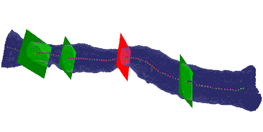
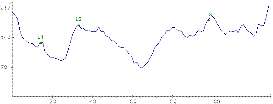
The segmented URT, its central path, and its cross--sectional profile at the three landmarks (vocal cords, caudal border of the cricoid cartilage, and cranial border of the sternum) and at the narrowest position (top); the line chart (bottom).
Assessment of Infrarenal Aortic Aneurysm
We used the cross-sectional profile in patients suffering from infra-renal aortic aneurysms (AAA). AAA are abnormal dilatations of the main arterial abdominal vessel due to atherosclerosis. AAA can be found in 2% of people older than 60 years. If the diameter is more than 5 cm than the person is at high risk for AAA rupture, which leads to death in 70-90%. For therapy two main options exist: surgery or endoluminal repair with stentgrafts. For optimal patient management the "true diameter" in 3D as well as the distance to the origin of the renal arteries (proximal aneurysma neck) as well as the extension to the iliac arteries (distal aneurysma neck) have to be known.
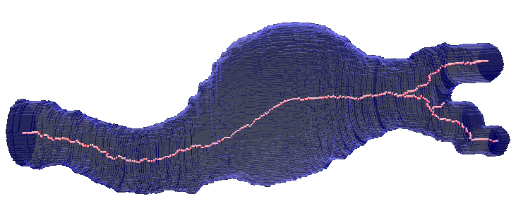
The segmented part of the infrarenal aorta and its central path. Along the central path the cross-sectional profile was computed. The following parameters could be derived from this approach: the maximum diameter in 3D as well as the length of the proximal and distal neck of the aneurysma. Since size of the aneurysma is regarded to be a prognosticated factor, the volume of the segmented aneurysma was determined too. At follow-up investigations the same parameters were derived.
Unravelling the Colon
Unravelling the colon is a new method to visualize the entire inner surface of the colon without the need for navigation. This is a minimally invasive technique that can be used for colorectal polyps and cancer detection. We have proposed an algorithm for unravelling the colon which is to digitally straighten and then flatten using reconstructed spiral/helical computer tomograph (CT) images. Comparing to virtual colonoscopy where polyps may be hidden from view behind the folds, the unravelled colon is more suitable for polyp detection, because the entire inner surface is displayed at one view.
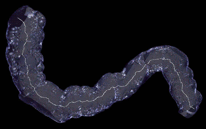
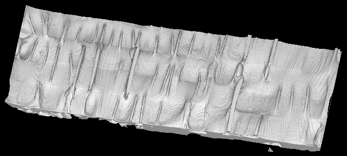
The segmented volume of a part of the cadavric phantom and its centerline (top) and the unravelled colon (bottom).
