CT view
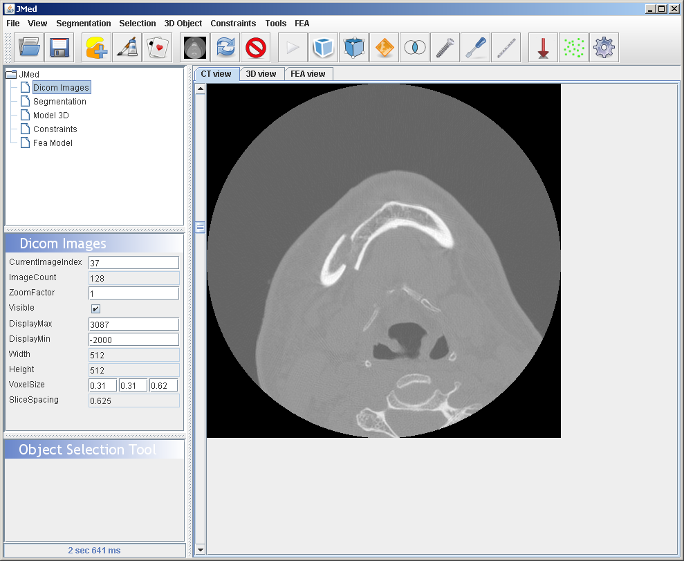
CT View tab.
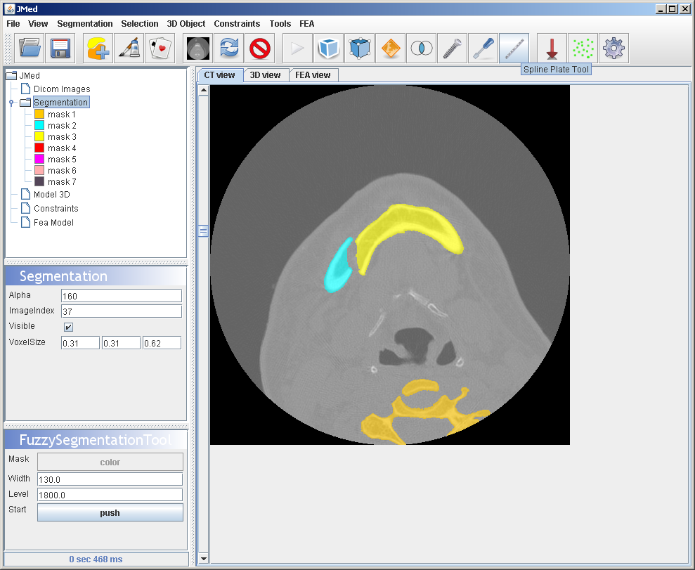
Segmentation is based on masks which are basically the individual bone fragments.
3D view
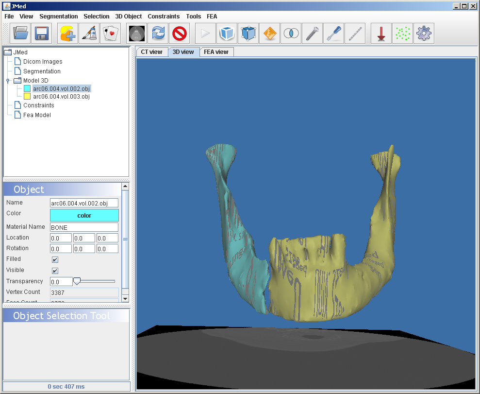
3D view also includes the CT slice in the Virtual World.
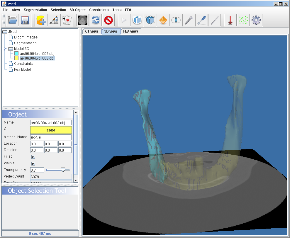
3D view. The cyan colored fragment is moved and rotated back to the position where it might have been before the fracture. Notice the difference to the CT image.
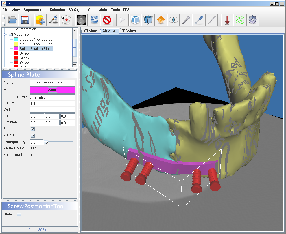
Implant insertion. Surgical plates and screws can be used for fixation.
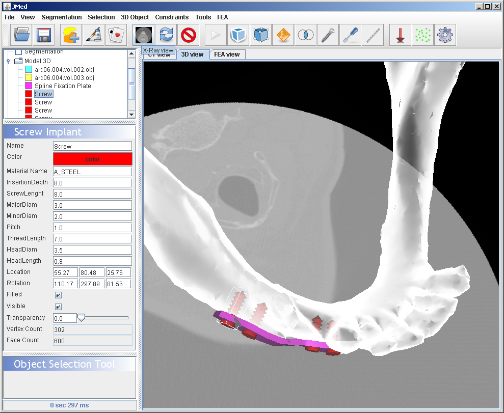
X-Ray view of the surgical plan. This view is a real time rendered X-ray simulation.
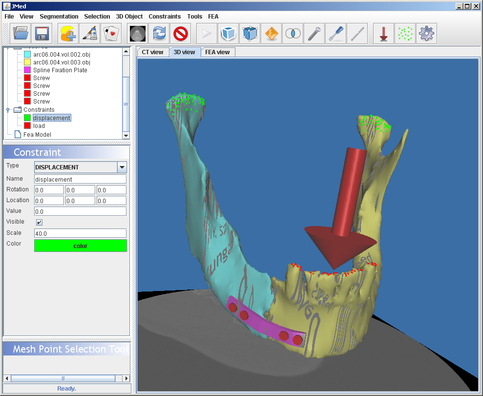
Mechanical model of the surgical plan. Green dots indicate the fix regions, and red dots indicate the region under load. The red arrow shows the direction of the load.
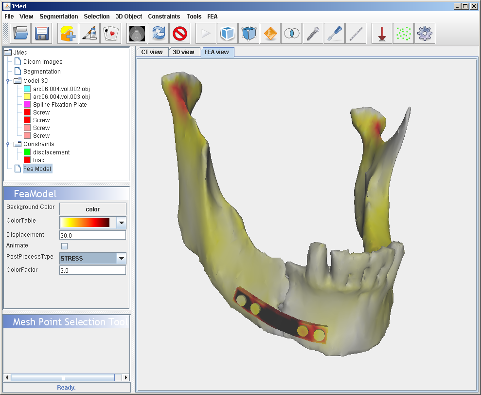
Result of the analysis.