The local organizers are:
- Péter Balázs,
- Antal Nagy,
- László Varga.
For further information please send email to wlst@inf.u-szeged.hu.
The local organizers are:
For further information please send email to wlst@inf.u-szeged.hu.
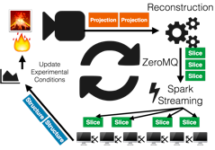
With every more efficient detectors, higher flux, and stable beamlines, comes the ability to probe time and length-scales previously unachievable. Of particular interest are the massive scale projects like the Human Brain Project, adult Zebra fish imaging, and dynamic imaging. All involve thousands to millions of measurements at the highest possible resolutions to cover mm to m length scales. The task of processing and analyzing such large collections of measurements is exceptionally difficult. We show how the commodity hardware-based ‘Big Data’ solutions can be adapted to the scientific domain to address the processing terabytes worth of measurements in a parallel, distributed manner. Building on the distributed frameworks of Apache Spark and Spark Imaging Layer, we have extended the common tomographic and image processing tools to work on these images enabling the use of many machines in parallel and drastically accelerating the speed and ease with which these large datasets can be stitched and analyzed. Our most recent developments enable the data to be analyzed and processed in real-time using the latest streaming and micro-batch processing techniques. Unique such an approach allows for fault-tolerant, distributed analytics to process complicated datasets and eventually provide feedback to both experimentalists and their equipment to allow for adaptation of measurements.
As new reconstruction methods are developed, they are often implemented in a scripting language (e.g. Matlab) for testing purposes. The application of these algorithms to large scale experimental data requires a time consuming re-implementation of the software in a high performance (parallel) programming language. With the use of the Spot matlab toolbox, the forward and backprojection operations of the ASTRA tomography toolbox (using the GPU) can be represented as a matrix. Therefore, advanced tomography algorithms using matrix-vector operations in Matlab can then directly be applied to large scale datasets. The use of these Spot operators is demonstrated with an implementation of a reconstruction method based on a non-convex data fit term which suppresses outliers in the projection data.
Optical diffraction tomography (ODT) is a non-invasive method, which enables quantitative measurement of three-dimensional, micrometre-sized elements that are transparent to visible light. The unique characteristics of this method make it a perfect tool for non-destructive characterization of such samples as optical elements, including fibre optics, and living biological specimens, e.g. singular biological cells and cell clusters. The principle of ODT is to capture a series of multiple holograms (optical wave’s measurements) that are obtained for various sample perspectives. Then, the holograms are numerical processed using a chosen tomographic reconstruction algorithm. The final result of ODT evaluation is a 3D reconstruction of refractive index, which give glimpse into internal structure of a sample and provides valuable information about its optical or/and biological properties.
Unfortunately, despite many great advantages of ODT, the method still suffers from a few fundamental problems. Most importantly, the ODT systems work with high-resolution and high-magnification microscope optics, which inherent characteristic is small depth of focus. This means that any inaccuracies in the sample rotation systems, which are used for alternation of measurement views, lead to displacement of a sample from the depth of focus and consequently results in blurring of sample projections. Recently, a holographic method has been reported, which avoids degradation of the tomographic data by utilizing an autofocus algorithm that allows for numerical correction of blur coming from defocusing of the sample projections. However, accuracy of the method depends on the object type and is usually far from satisfying. In this presentation, I will present a novel ODT solution, which addresses the problem of sample defocusing by employing structured illumination, i.e. simultaneous illumination form two highly off-axis directions. The method allows for accurate detection of the sample displacement by analysing interdependence of object waves corresponding to the individual illumination angles. Then, the obtained information is used for precise, numerical correction of defocusing. Moreover, application of structured illumination enables beating the fundamental theoretical limit related to the maximal achievable resolution and enlarge transfer function of the tomographic method even by a factor of two. The presented concept unlocks the full potential of ODT and can find application in measurement of very demanding samples such as photonics fibers and low-contrast objects.
Due to incompleteness of input data inherent to Limited Angle Tomography (LAT), specific additional constraints are usually employed to suppress image artifacts. In this work we demonstrate a two-stage regularization strategy dedicated to semi-piecewise-constant objects, named Total Variation Iterative Constraint (TVIC), successfully applied as a supplementary module for two different reconstruction algorithms: an X-ray type solver and a diffraction-wise solver. Numerical tests performed on a detailed phantom of a biological cell under conical illumination pattern show significant reduction of axial blurring in the reconstructed refractive index distribution after TVIC is added. Analogical results were reproduced with experimental data.
In Discrete Tomography, a main issue is to find conditions which ensure uniqueness of reconstruction in a given lattice grid A, by means of a finite set S of projections.
Several methods have been proposed in order to attack the problem, and theoretical results have been found, depending on the different employed discrete models.
In a previous paper, and in the context of binary tomography, we gave a necessary and sufficient uniqueness reconstruction condition, holding in the grid model for a set S consisting of four lattice directions. Starting from this result, we investigate two possible aspects which could be of interest in real applications.
On one hand we consider the tomographic reconstruction problem modeled as a linear system Wx=p, where x is the image to be reconstructed and p is the vector collecting the projections. We compute p for different sets S of four directions, pointing out how suitable choices allow perfect (noise-free) reconstructions. Further, in order to manage also nosy reconstructions, we propose to switch from the grid model to the strip model.
On the other hand, one could be prevented from using suitable sets of directions. Moreover, the tomographic problem is not necessary binary, and could involve gray-scale image reconstructions. In these cases a general uniqueness result is not known. However, it is often the case that one is not really interested in reconstructing the whole working grid A, but, rather, some portions of A which are of special interest for a given tomographic problem. To this, it is useful to know in advance the shape of the sub-regions of A where uniqueness can be quickly achieved.
A geometrical characterization of the uniqueness profile determined by a general set of two directions is presented, and examples are commented in view of possible applications.
With the latest developments in CMOS technology, it has been possible to exploit tomographic micros-copy at bright synchrotron facilities to unprecedented levels. We can routinely acquire 20 tomographic scans per second. The last hardware limitation in fast tomography has recently been solved at the Paul Scherrer Institut in Switzerland by introducing a new CMOS based detector that can stream data direct-ly to the server RAM instead of the conventional use of onboard camera RAM. Such a development puts tremendous pressure onto data handling as this system can acquire and stream continuously 7.7 BG/s directly to the server. In the meanwhile it opens new possibilities for data reduction schemes and even online 4D data analysis directly on the RAM of the server before images are saved to disk. The idea is to save only valuable data and filter out unnecessary projection images.
In Sweden the MAX IV facility is preparing for the future challenges in fast tomographic microscopy. I will present some plans and the search for sustainable solutions for handling Big Data with particular emphasis to in vivo imaging where radiation dose requirements trigger new developments in tomo-graphic reconstruction and quantification.
We discuss two new approaches for the selection of the regularization parameter in total variation (TV) regularized X-ray tomography. The first method is the S-curve method, which is based on a priori information about sparsity of the solution which may be available, for example, from an anatomical atlas. The parameter is selected to yield a priori expected level of sparsity of the solution. The second method is a multiresolution method which does not require a priori information. The approach is based on computing reconstructions at a few different resolutions and various values of regularization parameter. The chosen parameter is the smallest one resulting to approximately discretization-invariant TV norms of the reconstructions. The methods are tested with X-ray tomography data measured from a walnut specimen.
We consider the dynamic version of a major discrete tomography task: reconstruct the trajectories of moving particles from a small number of projections. The most relevant case in recent plasma physics applications involves projections from only two directions. It turns out that the computational complexity of these reconstruction tasks depends strongly on the type of prior information that is incorporated in the reconstruction. In this talk we present results that provide a particular sharp line between tractable (polynomial-time solvable) and intractable (NP-hard) problem instances. This is joint work with Peter Gritzmann (TU Muenchen).
X-ray computed tomography (CT) experiments performed at synchrotron radiation facilities require adequate computing and storage resources due to the large amount of acquired and reconstructed data produced. To satisfy the heterogeneous needs of beamline users, flexible solutions are also required. Moreover, the growing demand of quantitative image analysis impose an easy integration between the CT reconstruction process and the subsequent feature extraction step. This paper presents some of the software solutions adopted by the SYRMEP beamline of the Italian synchrotron radiation facility Elettra. By using the enhanced version of the reconstruction software here presented as well as data reduction and data analysis tools, beamline users can easily implement an integrated and comprehensive approach to the digital image processing and image analysis required by a tomography-oriented scientific workflow.
The increase of tomography data sizes is leading to an ever greater demand for parallelisation of reconstruction algorithms. The ASTRA Tomography Toolbox offers GPU-accelerated forward (FP) and backprojection (BP) operators for algorithm developers. In its latest open source release from December 2015, it also uses MPI to distribute computation over multiple servers and GPUs. It has a Python interface that lets algorithm developers use distributed versions of the FP and BP operators and also distribute their own Python code. In this talk, I will give an overview of the current features, and show how to use them for writing distributed reconstruction algorithms.
The talk will address some of the various CT techniques available at the ESRF: absorption tomography, phase contrast tomography (by propagation, grating interferometry or analyser based imaging), laminography, diffraction contrast tomography, XRD-CT and PDF-CT, as well as fluorescence tomography.
The application fields are as diverse as Materials and Life Science, Chemistry, Medicine, Cultural Heritage, Engineering, Palaeontology.
With the advent of beam nanofocusing devices (Kirkpatrick-Baez mirrors, CRLs, …) it is possible to raster scan the samples, and obtain space resolutions better than 50 nm. Conversely, owing to the high X-ray flux, sizeable samples like archaeological relics and paleontological findings can be scanned within a few hours at less than 50 μm resolution. Another challenging technique is time-resolved CT, with very fast detectors (DIMAX, Pilatus, …) that allow reconstructing an entire sample within a few seconds.
Typically, during one experiment as many as 10-20 TBytes of raw data can be produced. The requirements in terms of fast read-out pipelines and data storage are thus very stringent and will be also addressed. The reconstruction algorithms adopted are manifold (direct and algebraic methods) and tailored to tackle the different 3D imaging approaches and detection schemes.
For in-house research purposes, morphological and topological quantitative analyses of materials are also performed using the code iMorph.
Big Data is an emerging term covering various scenarios, use cases and activities that cannot be efficiently handled
using conventional approaches. While some of the related principles have already been in use for decades,
it is only recently that Big Data is gaining public recognition and popularity.
First, a quick overview will be presented about the motivation behind Big Data, including the principles and typical tasks involved. Special emphasis will be given to some of the challenges that one might encounter when facing Big Data problems.
Building on numerous best practices and experience gathered in the IT industry, a selection of tools and technologies
will be suggested that can help cope with Big Data problems. Furthermore, some examples will be shown for using these technologies in the realms of tomography.
Tomographic image reconstruction is an ill-posed inverse problem.
Bayesian approach can be used to propose acceptable solutions.
In this approach two main steps are important: choosing a prior model
and doing computations. In this work, we propose appropriate prior models which are rich via their hierarchical structures and appropriate associated algorithms which can be scalable for real 3D applications.
The new generation of imaging stations built at synchrotron light sources are able to reveal internal dynamics in fast processes and living organisms with high spatial and temporal resolution. But due to the ever increasing sampling rates they put strong demands on the complete signal processing chain. At KIT, we have developed hardware and software components covering the complete data life cycle from the sensor to the final storage at large-scale data facilities. Depending on the application a trade-off between image quality and processing performance of the image reconstruction has to be considered. Near real-time reconstruction is required to make control decisions and show first previews to the operator. For later analysis and visualization the image quality becomes imperative and quality-oriented algorithms have to be used. The ability to include a priori knowledge into a reconstruction model and produce a quality reconstruction from a lower number of projections is crucial for experiments with living organisms. These methods might prolong experiment duration by reducing the radiation dose inflicted for each frame. To handle all arising demands, we build a library of analytic and iterative reconstruction algorithms optimized for recent GPU architectures from AMD and NVIDIA. The throughput of the fastest algorithms exceeds 1 GB/s on a single GPU. We can execute the algorithms on a large-range of parallel computing hardware and schedule the workload across multiple parallel devices and nodes in the hybrid compute clusters. Close to linear scalability using up to 8 GPUs on a single computing node has been measured. In the talk, I will briefly describe the complete system design and, then, focus on image reconstruction and visualization sharing our experience on tuning the tomographic reconstruction for massively parallel architectures.
High resolution accurate tomography reconstruction requires supercomputer performance to provide results in reasonable time. Current GPUs can deliver such a performance if their special architectural features are taken into account during algorithmic design, i.e. the algorithm is decomposed to tens of thousand of independent computational threads that have no data dependent conditional statements and require neither synchronization nor communication. In this talk, we summarize the most important issues to achieve this goal and show solutions to simulate important phenomena in PET. The discussed approaches include the rewriting of transport equations for adjoint operators, the application of randomization, and combining different methods with multiple importance sampling.
ELI-ALPS will provide a wide range of atto-second pulses which will be used for performing chemical, biological, physical or medical experiments by international research groups. It is one pillar of the first international laser facility for the scientific user communities.
ELI-ALPS use the TANGO Controls framework to build up the central control system and to integrate the autonomous subsystems regarding monitoring and control. It will be also used for the implementation of some autonomous systems’ control system while others will be implemented differently. The central control system and the integration strategy of the autonomous systems is designed. Prototypes have been developed to clarify the requirements and to test the preliminary designs.
Online and offline data analysis as well as theoretical simulations will be executed in the CPU/GPU cluster of the facility. In case of very high demands external resources are planned to be allocated and used, supercomputer facilities and big data providers.