Jelenlegi hely
Retina image analysis
Chair of Pattern Recognition, University of Erlangen-Nuremberg (R. Bock, J. Meier, M. Mayer, J. Hornegger, G. Michelson)
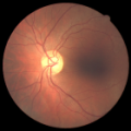
Glaucoma is one of the most common causes of blindness and it is becoming even more important considering the ageing society. Because healing of dead retinal nerve fibers is not possible, early detection and prevention is essential. Robust, automated mass-screening will help to extend the symptom-free life of affected patients.
This research project is part of the Collaborative Research Center 539 subproject A4. This research is mostly carried out at the Chair of Pattern Recognition, University of Erlangen-Nuremberg. L.G. Nyúl participated in this project during his Humboldt Research Fellowship year at the chair.
The main focus is the development of an automated, appearance based glaucoma classification system that does not depend on segmentation based measurements. Currently, we perform a purely data-driven approach that is applicable in large-scale screening examinations.
It applies a standard pattern recognition pipeline with a 2-stage classification step. Several types of image-based features were analyzed and are combined to capture glaucomatous structures. Certain disease independent variations such as illumination inhomogeneities, size differences, and vessel structures are eliminated in the preprocessing phase. The system currently achieves 86% success rate on a data set containing a mixture of 200 real images of healthy and glaucomatous eyes from the Erlangen Glaucoma Registry.
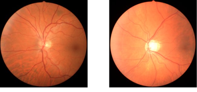
Figure 1: Fundus images acquired by Kowa NonMyd (45°). Left: Retinal image with pathologies. Right: Image with glaucomatous opticusatrophy.